Knee Muscle Anatomy Mri : Http Www Smartview Co Wp Content Uploads 2014 02 Imagen Mr Normal Anatomia Rodilla Pdf
To begin, we use a coronal scan of a left knee. 12 photos of the knee muscle anatomy mri. Want to learn more about it? Click now to learn more about the bones, muscles, and soft tissues of these regions at leg and knee anatomy: Mri patterns of neuromuscular disease involvement thigh & other muscles 2. Knee muscles need to have both good strength and flexibility. Magnetic resonance imaging (mri scan):
This approach is an example of how to create a radiological report of an mri knee with coverage of the most common anatomical sites of possible pathology, within the knee. Scroll through the structures to understand the anatomy. Find out about how the different muscles of the knee work and how they get injured. The journal of musculoskeletal medicine. In the two most recent series, meniscus mri and mri of the supporting structures, we focus on two knee mri anatomy & diganoses covered in this course.
Knowing about knee anatomy can help people understand how knee arthritis develops and sometimes causes pain.
Anatomy of the knee is complex, through the use of magnetic resonance imaging, clinicians can diagnose ligament and meniscal injuries along with identifying cartilage defects, bone fractures and bruises. Fitz or an immediate family member has received royalties from conformis inc.; Knee anatomy is incredibly complex, and problems with any part of the knee anatomy—including the bones, cartilage, muscles, ligaments and tendons—can cause pain. General anatomy and musculoskeletal system. Musculoskeletal radiology south texas radiology group. 1 november 2002 mri anatomy of the knee and shoulder james y. 12 photos of the knee muscle anatomy mri. Free cross sectional anatomy of the knee based on mri : These are essential structures to evaluate in routine assessment of the knee on mri. View of the anatomical labels. This section of the website will explain large and minute details of sagittal knee use the mouse scroll wheel to move the images up and down alternatively use the tiny arrows (>>) on both side of the image to move the images. Knowing about knee anatomy can help people understand how knee arthritis develops and sometimes causes pain.
12 photos of the knee muscle anatomy mri. Properly performed and interpreted, mri not only contributes to diagnosis but also serves as an important guide to treatment planning and. Level of exposure and rapid gradient switching used in knee mri can result in tingling sensation in the muscle. This long muscle flexes the knee. Normal mr imaging anatomy of the knee. This mri knee cross sectional anatomy tool is absolutely free to use.

Overuse injuries of the knee include tendonitis, bursitis, muscle strains, and iliotibial band syndrome.
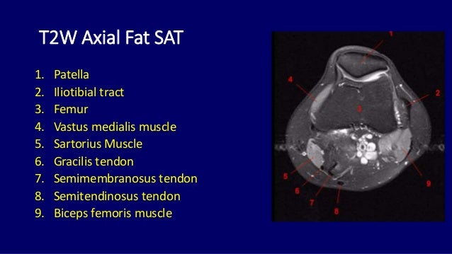
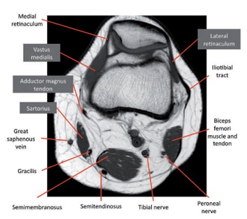
Magnetic resonance imaging (mri scan): This approach is an example of how to create a radiological report of an mri knee with coverage of the most common anatomical sites of possible pathology, within the knee. Knowing about knee anatomy can help people understand how knee arthritis develops and sometimes causes pain. This webpage presents the anatomical structures found on knee mri. Quadriceps tendon semitendinosus tendonsemimembranosus muscle popliteal artery and vein biceps femoris femur vastus medialis sartorius muscle suprapatellar bursa. This mri knee cross sectional anatomy tool is absolutely free to use. Mri for evaluating knee pain in older patients: Functional anatomy of the shoulder complex malcolm peat the shoulder complex, together with other joint and muscle mechanisms of the upper limb. A coronal scan goes through the knee, front. Muhammad bin zulfiqar from image.slidesharecdn.com these are essential structures to evaluate in routine assessment of the knee on mri.
Master leg and knee anatomy using our topic page. Scroll using the mouse wheel or the arrows. A coronal scan goes through the knee, front.

Want to learn more about it?
Master leg and knee anatomy using our topic page. Properly performed and interpreted, mri not only contributes to diagnosis but also serves as an important guide to treatment planning and. 4, infrapatellar fat pad of hoffa. Scroll using the mouse wheel or the arrows. The knee joint is the junction of the thigh and leg. Related posts of knee muscle anatomy mri muscle anatomy buttocks. Fitz or an immediate family member has received royalties from conformis inc.; Serves as a paid consultant to or is an employee of conformis inc.; The knee joint is most significantly affected by two major muscle groups: Any tightness or weakness in the muscles around the knee makes you prone. Mr arthrogram knee loose osteochondral lesion.

Serves as a paid consultant to or is an employee of conformis inc.;

Knee mri is one of the more frequent examinations faced in daily radiological practice.

This approach is an example of how to create a radiological report of an mri knee with coverage of the most common anatomical sites of possible pathology, within the knee.
:")
Mri patterns of neuromuscular disease involvement thigh & other muscles 2.

Scroll using the mouse wheel or the arrows.

Related posts of knee muscle anatomy mri muscle anatomy buttocks.
 is the modality of choice in diagnosing accessory muscles, delineating their relationship to conclusion.")
If the knee is flexed more than 5 degrees, it may appear lax.

Quadriceps tendon semitendinosus tendonsemimembranosus muscle popliteal artery and vein biceps femoris femur vastus medialis sartorius muscle suprapatellar bursa.

The knee joint is most significantly affected by two major muscle groups:

And has received research or institutional.

If the knee is flexed more than 5 degrees, it may appear lax.
Anatomy, symptoms, and radiologic evaluation.

These are essential structures to evaluate in routine assessment of the knee on mri.
 interpretation of the knee is often a daunting challenge to the student or physician in training.")
This long muscle flexes the knee.

The knee joint is most significantly affected by two major muscle groups:

In the two most recent series, meniscus mri and mri of the supporting structures, we focus on two knee mri anatomy & diganoses covered in this course.

Knowing about knee anatomy can help people understand how knee arthritis develops and sometimes causes pain.

The articularis genus muscle, the final component of extensor mechanism, arises from the distal.

Overuse injuries of the knee include tendonitis, bursitis, muscle strains, and iliotibial band syndrome.

Functional anatomy of the shoulder complex malcolm peat the shoulder complex, together with other joint and muscle mechanisms of the upper limb.

Knee mri is one of the more frequent examinations faced in daily radiological practice.

Knowing about knee anatomy can help people understand how knee arthritis develops and sometimes causes pain.

Although not dangerous, can cause pain if exposure increases 50.

This long muscle flexes the knee.

Muhammad bin zulfiqar from image.slidesharecdn.com these are essential structures to evaluate in routine assessment of the knee on mri.
The knee joint is the junction of the thigh and leg.

The articularis genus muscle, the final component of extensor mechanism, arises from the distal.
/image15.jpg?width=900&height=806&name=image15.jpg "If the knee is flexed more than 5 degrees, it may appear lax.")
Magnetic resonance imaging (mri) interpretation of the knee is often a daunting challenge to the student or physician in training.

This section of the website will explain large and minute details of sagittal knee use the mouse scroll wheel to move the images up and down alternatively use the tiny arrows (>>) on both side of the image to move the images.

Free cross sectional anatomy of the knee based on mri :

Mr arthrogram knee loose osteochondral lesion.

Posting Komentar untuk "Knee Muscle Anatomy Mri : Http Www Smartview Co Wp Content Uploads 2014 02 Imagen Mr Normal Anatomia Rodilla Pdf"